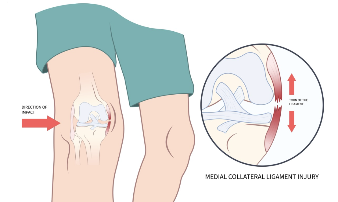
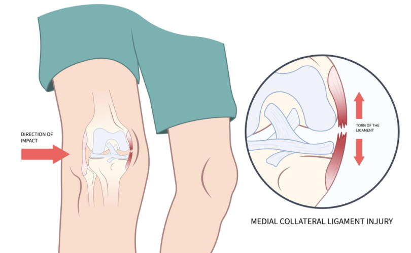
The MCL is a commonly injured ligament on the inside of the knee. This ligament is structurally important for the stability of the knee, and critical for the performance of both sport and physical activity, as well as everyday tasks and functions.
Anatomy
The knee is a hinge joint between the femur (thigh bone), tibia (shin bone), fibula (bone on the outside of the shin), and patella (kneecap). There are four main ligaments around and within the knee that are most important for maintaining stability of the knee. These include the ACL and PCL within the knee joint itself, and the two collateral ligaments on the sides of the knee.
The Medial Collateral Ligament (MCL) and Lateral Collateral Ligament (LCL) are large ligaments on the inside and outside of the knee respectively. They prevent the knee from bending sideways too much in either direction.
The MCL joins the femur (thigh) to the tibia (shin) on the inside of the knee. It aims to prevent the knee bending inwards, and the knee joint gapping too far on the medial (inner) side.
Mechanism of Injury
The MCL can sustain injury from:
- Direct impact to the outside of the leg. When this causes a valgus force to the knee (ie the knee bends inwards), it can stress and ultimately sprain or tear the MCL.
- Non-contact: twisting/pivoting with the foot in contact with the ground can result in the same valgus force on the knee.
While these mechanisms of injury are often present on the sporting field, these same pivoting and twisting forces can be a common occurrence at home and in everyday life.
Common Associated Injuries
- ACL
- Meniscus
Presentation
Some of the common characteristics of an MCL injury include:
- Mechanism of injury as described above.
- Pain on the inner knee, including tenderness on palpation.
- Possible popping sound at time of injury.
- Swelling localised to the inside of the knee.
- Limping, which worsens with higher grade tears.
- Knee feeling unstable or giving way.
Medial Stress Test: the medial or valgus stress test is used to ascertain whether the is a likelihood of injury to the MCL, and if so, how severe that injury may be.
Grading
Grade 1: generally mild pain, small amount of swelling, and pain on a medial stress test without laxity.
Grade 2: increased pain and swelling, with pain and laxity present on a medial stress test.
Grade 3: severe pain and swelling, with pain* and extreme laxity on medial stress test and often feelings of instability or giving way while attempting to walk.
*Note: in some cases, Grade 3 tears can have less pain on stress test.
Treatment
In most cases of Grade 1-2 MCL tears, non-surgical rehabilitation is considered ideal, and our physiotherapists can assist you proficiently with that. Here is a general outline of how that looks:
- Protection, Healing, Swelling Control: Initially, the ligament must be protected. For Grade 1 injuries, this may just look like rest and swelling control.
However for Grade 2-3 tears, this generally involves a locked ROM brace. This is a brace that restricts the amount a knee can bend and straighten, in order to keep the ligament in a shortened position to encourage tissue healing. - Range of Motion (ROM): the next thing to be considered is maintaining, or achieving full ROM, with exercise and manual therapy from a physiotherapist.
- Strength/Stability: once the knee can tolerate it, strengthening can occur of all the muscles around the knee, as well as improving balance and stability.
- Function: eventually, there is a return to functional movement, such as running or training, and this includes exercises to increase proprioception and work safely at speed.
- Return to Sport (or full function): Finally, the physiotherapist will provide criteria to be discharged from physiotherapy and return to full activities without restriction. This will be sport-specific and catered to the individual.
In some cases of Grade 2-3 injuries to the MCL, surgery is advisable, and particularly if there is other associated injury in the knee. In that case, a physiotherapist or GP may advise seeing a specialist, and then rehabilitation post-surgery can be commenced with your physiotherapist.
If you have medial knee pain or any acute knee injury, we’d love to hear about it and see you for an assessment! We can be your first stop for advice as to whether you can immediately begin rehabilitation, if a brace is indicated, and whether there is a need to be seeking imaging and a specialist review.
Written by Joshua Round, Physiotherapist at Prime Physiotherapy.